

Description
Primary ciliary dyskinesia is a disorder that is characterized by chronic respiratory tract infections, abnormally positioned internal organs, and difficulties having biological children (decreased fertility). The signs and symptoms of this condition are caused by abnormal cilia and flagella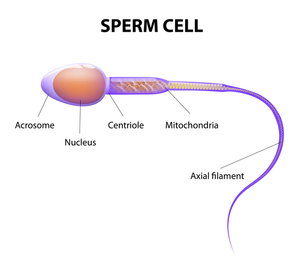 . Cilia are microscopic, finger-like projections that stick out from the surface of cells. Cilia help cells move where they are needed; they also help move substances within the body. Flagella, which are similar to cilia, are tail-like structures that propel sperm cells forward.
Without properly functioning cilia, people with primary ciliary dyskinesia often have problems removing fluid and particles from their airways. Most babies with primary ciliary dyskinesia experience breathing problems at birth (neonatal respiratory distress), which suggests that cilia also play an important role in clearing fetal fluid from the lungs. Children with primary ciliary dyskinesia typically have year-round nasal congestion and a chronic cough beginning in the first year of life. Because affected individuals also have trouble removing bacteria from the respiratory tract, they may experience frequent respiratory tract infections beginning in early childhood. Chronic respiratory tract infections can result in a condition called bronchiectasis, which damages the passages that lead from the windpipe to the lungs (bronchi). Bronchiectasis can cause life-threatening breathing problems.
Another feature of primary ciliary dyskinesia is recurrent ear infections (otitis media), especially in young children. Otitis media can lead to permanent hearing loss if left untreated. These ear infections are likely related to abnormal cilia within the inner ear.
About 40 percent of people with primary ciliary dyskinesia have a mirror-image reversal of their internal organs (situs inversus totalis). For example, the heart is on the right side of the body instead of the left in these individuals. These abnormalities arise early in embryonic development when the differences between the left and right sides of the body are established. Situs inversus totalis does not typically cause additional health problems. When someone with primary ciliary dyskinesia has situs inversus totalis, they are often said to have Kartagener syndrome.
Heterotaxy syndrome (sometimes also called situs ambiguous) is another disorder of organ development that can be associated with primary ciliary dyskinesia. Approximately 9 to 12 percent of people with primary ciliary dyskinesia have heterotaxy syndrome, which is characterized by abnormalities of the heart, liver, intestines, or spleen. These organs may be structurally abnormal or improperly positioned. In addition, affected individuals may lack a spleen (asplenia) or have multiple spleens (polysplenia). Heterotaxy syndrome is also a result of problems establishing the left and right sides of the body during embryonic development. The severity of heterotaxy syndrome varies widely among affected individuals, and people with this condition may have heart abnormalities that can be life-threatening.
Primary ciliary dyskinesia can also cause fertility problems. Vigorous movements of the flagella are necessary to propel the sperm cells forward to the egg cell. Because their sperm do not move properly, males with primary ciliary dyskinesia often have decreased fertility. Decreased fertility also occurs in some affected females and is likely due to abnormal cilia in the fallopian tubes, which impairs the movement of the egg cell from the ovary to the uterus.
In rare cases, individuals with primary ciliary dyskinesia have an accumulation of fluid in the brain (hydrocephalus). Researchers do not fully understand why some people with primary ciliary dyskinesia develop hydrocephalus.
Frequency
It is estimated that as many as 1 in 7,500 people worldwide have primary ciliary dyskinesia.
Causes
Variants (also called mutations) in one of over 50 different genes can cause primary ciliary dyskinesia. Many of these genes provide instructions for making proteins that form the inner structure of cilia and produce the force needed for cilia to bend. Coordinated back and forth movement of cilia is necessary for the normal functioning of many organs and tissues. The movement of cilia also supports cell transport and helps establish the left-right axis (the imaginary line that separates the left and right sides of the body) during embryonic development.
Many of the variants that cause primary ciliary dyskinesia disrupt the coordinated movement of cilia. Because cilia have many important functions within the body, abnormalities in the structure or function of cilia can cause a variety of signs and symptoms.
Variants in the DNAH5 and DNAH11 genes account for approximately 33 percent of all cases of primary ciliary dyskinesia. These genes provide instructions for producing proteins that are part of a larger group of proteins (a complex) called dynein. Dynein makes up the structures (arms) within the cilia that generate the force needed for cilia to move.
Variants in the CCDC40 gene account for approximately 9 percent of cases of primary ciliary dyskinesia. This gene provides instructions for producing a protein that helps with the proper assembly of dynein arms. This protein also helps assemble a protein complex that regulates the movement of the dynein arms.
Variants in each of the other genes associated with this condition are found in only a small percentage of cases. In 20 to 30 percent of people with primary ciliary dyskinesia, the cause of the disorder is unknown.
Inheritance
Primary ciliary dyskinesia is typically inherited in an autosomal recessive pattern , which means both copies of the gene in each cell must have a variant to cause the disorder. The parents of an individual with an autosomal recessive condition each carry one copy of the altered gene, but they typically do not show signs and symptoms of the condition.
In some cases, primary ciliary dyskinesia is inherited in an X-linked pattern. A condition is considered to follow an X-linked pattern if the altered gene that causes the disorder is located on the X chromosome, one of the two sex chromosomes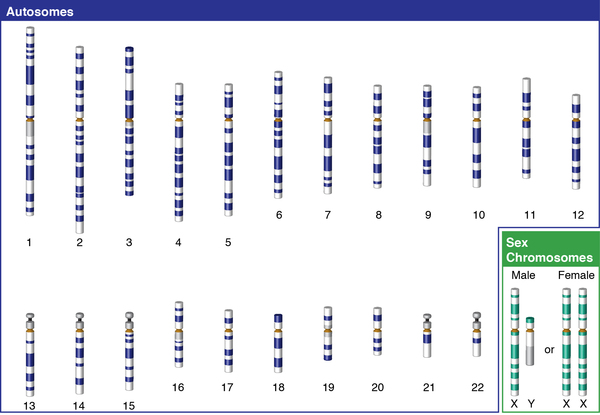 in each cell. In males (who have only one X chromosome) a variant in the only copy of the gene in each cell is sufficient to cause the condition. In females (who have two copies of the X chromosome) one altered copy of the gene may or may not cause the features of the condition.
In rare cases, primary ciliary dyskinesia is inherited in an autosomal dominant pattern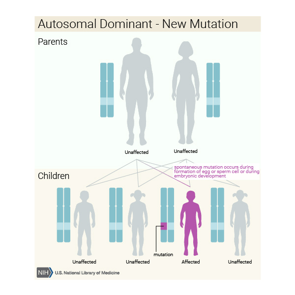 , which means one copy of the altered gene in each cell is sufficient to cause the disorder. In most of these cases, the condition is a result of new (de novo) variants in the gene that occur during the formation of reproductive cells (eggs or sperm) in an affected individual's parent or during early embryonic development. These affected individuals typically have no history of the disorder in their family.
Other Names for This Condition
- CILD
- Kartagener syndrome
- PCD
Additional Information & Resources
Genetic Testing Information
Genetic and Rare Diseases Information Center
Patient Support and Advocacy Resources
Clinical Trials
Catalog of Genes and Diseases from OMIM
- CILIARY DYSKINESIA, PRIMARY, 10; CILD10
- CILIARY DYSKINESIA, PRIMARY, 11; CILD11
- CILIARY DYSKINESIA, PRIMARY, 12; CILD12
- CILIARY DYSKINESIA, PRIMARY, 13; CILD13
- CILIARY DYSKINESIA, PRIMARY, 14; CILD14
- CILIARY DYSKINESIA, PRIMARY, 15; CILD15
- CILIARY DYSKINESIA, PRIMARY, 16; CILD16
- CILIARY DYSKINESIA, PRIMARY, 17; CILD17
- CILIARY DYSKINESIA, PRIMARY, 18; CILD18
- CILIARY DYSKINESIA, PRIMARY, 19; CILD19
- CILIARY DYSKINESIA, PRIMARY, 1; CILD1
- CILIARY DYSKINESIA, PRIMARY, 20; CILD20
- CILIARY DYSKINESIA, PRIMARY, 21; CILD21
- CILIARY DYSKINESIA, PRIMARY, 22; CILD22
- CILIARY DYSKINESIA, PRIMARY, 23; CILD23
- CILIARY DYSKINESIA, PRIMARY, 24; CILD24
- CILIARY DYSKINESIA, PRIMARY, 25; CILD25
- CILIARY DYSKINESIA, PRIMARY, 26; CILD26
- CILIARY DYSKINESIA, PRIMARY, 27; CILD27
- CILIARY DYSKINESIA, PRIMARY, 28; CILD28
- CILIARY DYSKINESIA, PRIMARY, 29; CILD29
- CILIARY DYSKINESIA, PRIMARY, 2; CILD2
- CILIARY DYSKINESIA, PRIMARY, 30; CILD30
- CILIARY DYSKINESIA, PRIMARY, 32; CILD32
- CILIARY DYSKINESIA, PRIMARY, 33; CILD33
- CILIARY DYSKINESIA, PRIMARY, 34; CILD34
- CILIARY DYSKINESIA, PRIMARY, 35; CILD35
- CILIARY DYSKINESIA, PRIMARY, 36, X-LINKED; CILD36
- CILIARY DYSKINESIA, PRIMARY, 37; CILD37
- CILIARY DYSKINESIA, PRIMARY, 38; CILD38
- CILIARY DYSKINESIA, PRIMARY, 39; CILD39
- CILIARY DYSKINESIA, PRIMARY, 3; CILD3
- CILIARY DYSKINESIA, PRIMARY, 40; CILD40
- CILIARY DYSKINESIA, PRIMARY, 41; CILD41
- CILIARY DYSKINESIA, PRIMARY, 42; CILD42
- CILIARY DYSKINESIA, PRIMARY, 43; CILD43
- CILIARY DYSKINESIA, PRIMARY, 44; CILD44
- CILIARY DYSKINESIA, PRIMARY, 45; CILD45
- CILIARY DYSKINESIA, PRIMARY, 46; CILD46
- CILIARY DYSKINESIA, PRIMARY, 47, AND LISSENCEPHALY; CILD47
- CILIARY DYSKINESIA, PRIMARY, 48, WITHOUT SITUS INVERSUS; CILD48
- CILIARY DYSKINESIA, PRIMARY, 49, WITHOUT SITUS INVERSUS; CILD49
- CILIARY DYSKINESIA, PRIMARY, 4; CILD4
- CILIARY DYSKINESIA, PRIMARY, 50; CILD50
- CILIARY DYSKINESIA, PRIMARY, 51; CILD51
- CILIARY DYSKINESIA, PRIMARY, 52; CILD52
- CILIARY DYSKINESIA, PRIMARY, 53; CILD53
- CILIARY DYSKINESIA, PRIMARY, 54; CILD54
- CILIARY DYSKINESIA, PRIMARY, 55; CILD55
- CILIARY DYSKINESIA, PRIMARY, 5; CILD5
- CILIARY DYSKINESIA, PRIMARY, 6; CILD6
- CILIARY DYSKINESIA, PRIMARY, 7; CILD7
- CILIARY DYSKINESIA, PRIMARY, 8; CILD8
- CILIARY DYSKINESIA, PRIMARY, 9; CILD9
Scientific Articles on PubMed
References
- Despotes KA, Zariwala MA, Davis SD, Ferkol TW. Primary Ciliary Dyskinesia: A Clinical Review. Cells. 2024 Jun 4;13(11):974. doi: 10.3390/cells13110974. Citation on PubMed
- Escudier E, Duquesnoy P, Papon JF, Amselem S. Ciliary defects and genetics of primary ciliary dyskinesia. Paediatr Respir Rev. 2009 Jun;10(2):51-4. doi: 10.1016/j.prrv.2009.02.001. Epub 2009 Apr 18. Citation on PubMed
- Failly M, Bartoloni L, Letourneau A, Munoz A, Falconnet E, Rossier C, de Santi MM, Santamaria F, Sacco O, DeLozier-Blanchet CD, Lazor R, Blouin JL. Mutations in DNAH5 account for only 15% of a non-preselected cohort of patients with primary ciliary dyskinesia. J Med Genet. 2009 Apr;46(4):281-6. doi: 10.1136/jmg.2008.061176. Citation on PubMed
- Failly M, Saitta A, Munoz A, Falconnet E, Rossier C, Santamaria F, de Santi MM, Lazor R, DeLozier-Blanchet CD, Bartoloni L, Blouin JL. DNAI1 mutations explain only 2% of primary ciliary dykinesia. Respiration. 2008;76(2):198-204. doi: 10.1159/000128567. Epub 2008 Apr 23. Citation on PubMed
- Hannah WB, Seifert BA, Truty R, Zariwala MA, Ameel K, Zhao Y, Nykamp K, Gaston B. The global prevalence and ethnic heterogeneity of primary ciliary dyskinesia gene variants: a genetic database analysis. Lancet Respir Med. 2022 May;10(5):459-468. doi: 10.1016/S2213-2600(21)00453-7. Epub 2022 Jan 17. Citation on PubMed
- Horani A, Brody SL, Ferkol TW. Picking up speed: advances in the genetics of primary ciliary dyskinesia. Pediatr Res. 2014 Jan;75(1-2):158-64. doi: 10.1038/pr.2013.200. Epub 2013 Nov 5. Citation on PubMed or Free article on PubMed Central
- Hornef N, Olbrich H, Horvath J, Zariwala MA, Fliegauf M, Loges NT, Wildhaber J, Noone PG, Kennedy M, Antonarakis SE, Blouin JL, Bartoloni L, Nusslein T, Ahrens P, Griese M, Kuhl H, Sudbrak R, Knowles MR, Reinhardt R, Omran H. DNAH5 mutations are a common cause of primary ciliary dyskinesia with outer dynein arm defects. Am J Respir Crit Care Med. 2006 Jul 15;174(2):120-6. doi: 10.1164/rccm.200601-084OC. Epub 2006 Apr 20. Citation on PubMed or Free article on PubMed Central
- Kennedy MP, Omran H, Leigh MW, Dell S, Morgan L, Molina PL, Robinson BV, Minnix SL, Olbrich H, Severin T, Ahrens P, Lange L, Morillas HN, Noone PG, Zariwala MA, Knowles MR. Congenital heart disease and other heterotaxic defects in a large cohort of patients with primary ciliary dyskinesia. Circulation. 2007 Jun 5;115(22):2814-21. doi: 10.1161/CIRCULATIONAHA.106.649038. Epub 2007 May 21. Citation on PubMed
- Leigh MW, Pittman JE, Carson JL, Ferkol TW, Dell SD, Davis SD, Knowles MR, Zariwala MA. Clinical and genetic aspects of primary ciliary dyskinesia/Kartagener syndrome. Genet Med. 2009 Jul;11(7):473-87. doi: 10.1097/GIM.0b013e3181a53562. Citation on PubMed or Free article on PubMed Central
- Morillas HN, Zariwala M, Knowles MR. Genetic causes of bronchiectasis: primary ciliary dyskinesia. Respiration. 2007;74(3):252-63. doi: 10.1159/000101783. Citation on PubMed
- Raidt J, Riepenhausen S, Pennekamp P, Olbrich H, Amirav I, Athanazio RA, Aviram M, Balinotti JE, Bar-On O, Bode SFN, Boon M, Borrelli M, Carr SB, Crowley S, Dehlink E, Diepenhorst S, Durdik P, Dworniczak B, Emiralioglu N, Erdem E, Fonnesu R, Gracci S, Grosse-Onnebrink J, Gwozdziewicz K, Haarman EG, Hansen CR, Hogg C, Holgersen MG, Kerem E, Korner RW, Kotz K, Kouis P, Loebinger MR, Lorent N, Lucas JS, Maj D, Mall MA, Marthin JK, Martinu V, Mazurek H, Mitchison HM, Nothe-Menchen T, Ozcelik U, Pifferi M, Pogorzelski A, Ringshausen FC, Roehmel JF, Rovira-Amigo S, Rumman N, Schlegtendal A, Shoemark A, Sperstad Kennelly S, Staar BO, Sutharsan S, Thomas S, Ullmann N, Varghese J, von Hardenberg S, Walker WT, Wetzke M, Witt M, Yiallouros P, Zschocke A, Zietkiewicz E, Nielsen KG, Omran H. Analyses of 1236 genotyped primary ciliary dyskinesia individuals identify regional clusters of distinct DNA variants and significant genotype-phenotype correlations. Eur Respir J. 2024 Aug 8;64(2):2301769. doi: 10.1183/13993003.01769-2023. Print 2024 Aug. Citation on PubMed
- Shapiro AJ, Davis SD, Ferkol T, Dell SD, Rosenfeld M, Olivier KN, Sagel SD, Milla C, Zariwala MA, Wolf W, Carson JL, Hazucha MJ, Burns K, Robinson B, Knowles MR, Leigh MW; Genetic Disorders of Mucociliary Clearance Consortium. Laterality defects other than situs inversus totalis in primary ciliary dyskinesia: insights into situs ambiguus and heterotaxy. Chest. 2014 Nov;146(5):1176-1186. doi: 10.1378/chest.13-1704. Citation on PubMed or Free article on PubMed Central
- Sutherland MJ, Ware SM. Disorders of left-right asymmetry: heterotaxy and situs inversus. Am J Med Genet C Semin Med Genet. 2009 Nov 15;151C(4):307-17. doi: 10.1002/ajmg.c.30228. Citation on PubMed
- Wee WB, Kinghorn B, Davis SD, Ferkol TW, Shapiro AJ. Primary Ciliary Dyskinesia. Pediatrics. 2024 Jun 1;153(6):e2023063064. doi: 10.1542/peds.2023-063064. Citation on PubMed
- Zariwala MA, Despotes KA, Davis SD. Primary Ciliary Dyskinesia. 2007 Jan 24 [updated 2025 May 22]. In: Adam MP, Bick S, Mirzaa GM, Pagon RA, Wallace SE, Amemiya A, editors. GeneReviews(R) [Internet]. Seattle (WA): University of Washington, Seattle; 1993-2026. Available from http://www.ncbi.nlm.nih.gov/books/NBK1122/ Citation on PubMed
The information on this site should not be used as a substitute for professional medical care or advice. Contact a health care provider if you have questions about your health.