

Description
Recurrent hydatidiform mole is a condition that affects women and is characterized by the occurrence of at least two abnormal pregnancies that result in the formation of hydatidiform moles. A hydatidiform mole is a mass that forms early in pregnancy and is made up of cells from an abnormally developed embryo and placenta. Normally, the embryo would develop into a fetus and the placenta would grow to provide nutrients to the growing fetus. When a hydatidiform mole occurs once, it is known as sporadic hydatidiform mole; if it happens again, the condition is known as recurrent hydatidiform mole.
The first symptom of a hydatidiform mole is often vaginal bleeding in the first trimester of pregnancy. During an ultrasound examination, the abnormal placenta appears as numerous small sacs, often described as resembling a bunch of grapes.
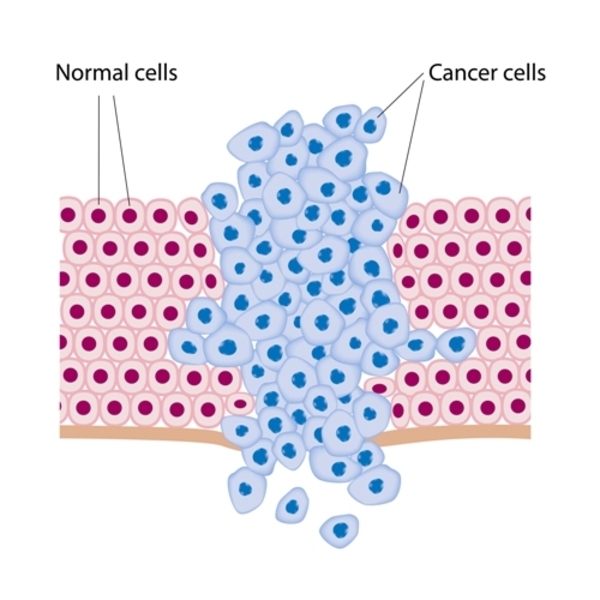
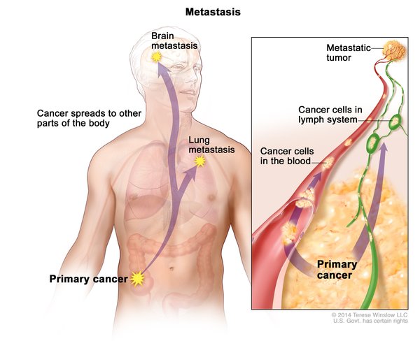
Hydatidiform moles are not naturally discharged from the body and must be surgically removed, typically by the end of the first trimester. After removal, there is up to a 20 percent risk that any tissue left behind will continue to grow and become a cancerous (malignant) tumor called a persistent mole. If the tumor invades the surrounding tissue of the uterus, it is called an invasive mole. In rare cases, this malignant tumor can transform into a different form of cancer called gestational choriocarcinoma that can spread (metastasize) to other tissues such as the liver, lungs, or brain.
Frequency
Hydatidiform moles occur in 1 in 600 to 1,000 pregnancies in western countries. One to six percent of previously affected women will have a recurrent hydatidiform mole. Gestational choriocarcinoma occurs in 1 in 20,000 to 50,000 pregnancies in the United States.
Causes
Mutations in multiple genes have been found to cause recurrent hydatidiform mole. About 55 percent of cases of this condition are caused by NLRP7 gene mutations and about 5 percent of cases are caused by KHDC3L gene mutations. Mutations in other genes each account for a small percentage of cases.
The proteins produced from the NLRP7 and KHDC3L genes are critical for normal egg cell (oocyte) development, which impacts embryonic development. Within oocytes, the exact role of NLRP7 and KHDC3L proteins are not known. However, they are thought to play a role in a phenomenon known as genomic imprinting. Through genomic imprinting certain genes are turned off (inactivated) based on which parent the copy of the gene came from. For most genes, both copies of the gene (one copy inherited from each parent) are active in all cells. However, for a small subset of genes, only one of the two copies is active and the other is turned off. For some of these genes, the copy from the father is normally active, while for others, the copy from the mother is normally active.
NLRP7 or KHDC3L gene mutations result in the production of proteins with impaired function. As a result, oocytes do not develop normally. A pregnancy that results from an abnormal oocyte cannot develop properly, resulting in recurrent hydatidiform mole. NLRP7 or KHDC3L gene mutations can also prevent proper imprinting of multiple genes that contribute to a developing embryo, leading to abnormal gene activity (expression). It is not clear if problems with imprinting also contribute to the development of a hydatidiform mole. In women with NLRP7 or KHDC3L gene mutations, a hydatidiform mole will develop in every pregnancy that occurs with her egg cells.
A small number of cases of recurrent hydatidiform mole have been found to be caused by mutations in genes that play important roles in the production of oocytes and sperm cells. The proteins produced from these genes are involved in the normal process of exchanging genetic material between chromosomes in preparation for cell division during oocyte and sperm cell production. These proteins are needed to make breaks in the chromosomes so that genetic information can be exchanged.

Mutations in these genes prevent the normal function of the proteins involved in the exchange of genetic material. Without the exchange of genetic material, cell division is often stopped. In affected women, this can lead to the production of abnormal oocytes that do not contain chromosomes. When a normal sperm cell fertilizes one of these oocytes, the resulting embryo has only one set of chromosomes. Because the embryo has no genes from the mother, the pregnancy cannot develop normally, resulting in a hydatidiform mole. In women with these rare gene mutations, every pregnancy that occurs with her egg cells will result in a hydatidiform mole or pregnancy loss (miscarriage).
In some cases of recurrent hydatidiform mole, no mutations in any of the genes associated with the condition have been identified. In these instances, the cause of the condition is unknown.
Inheritance
Recurrent hydatidiform mole is inherited in an autosomal recessive pattern, which means both copies of the gene in each cell have mutations. The parents of an individual with an autosomal recessive condition each carry one copy of the mutated gene, but they typically do not show signs and symptoms of the condition. Recurrent hydatidiform mole seems to have an autosomal recessive inheritance pattern even when the genetic cause of the condition is unknown.

Other Names for This Condition
- Familial recurrent hydatidiform mole
- FRHM
- Recurrent androgenetic hydatidiform mole
- Recurrent biparental hydatidiform mole
Additional Information & Resources
Genetic Testing Information
Genetic and Rare Diseases Information Center
Patient Support and Advocacy Resources
Catalog of Genes and Diseases from OMIM
Scientific Articles on PubMed
References
- Fallahian M, Sebire NJ, Savage PM, Seckl MJ, Fisher RA. Mutations in NLRP7 and KHDC3L confer a complete hydatidiform mole phenotype on digynic triploid conceptions. Hum Mutat. 2013 Feb;34(2):301-8. doi: 10.1002/humu.22228. Epub 2012 Nov 2. Citation on PubMed
- Hayward BE, De Vos M, Talati N, Abdollahi MR, Taylor GR, Meyer E, Williams D, Maher ER, Setna F, Nazir K, Hussaini S, Jafri H, Rashid Y, Sheridan E, Bonthron DT. Genetic and epigenetic analysis of recurrent hydatidiform mole. Hum Mutat. 2009 May;30(5):E629-39. doi: 10.1002/humu.20993. Citation on PubMed
- Hui P, Buza N, Murphy KM, Ronnett BM. Hydatidiform Moles: Genetic Basis and Precision Diagnosis. Annu Rev Pathol. 2017 Jan 24;12:449-485. doi: 10.1146/annurev-pathol-052016-100237. Citation on PubMed
- Mahadevan S, Wen S, Wan YW, Peng HH, Otta S, Liu Z, Iacovino M, Mahen EM, Kyba M, Sadikovic B, Van den Veyver IB. NLRP7 affects trophoblast lineage differentiation, binds to overexpressed YY1 and alters CpG methylation. Hum Mol Genet. 2014 Feb 1;23(3):706-16. doi: 10.1093/hmg/ddt457. Epub 2013 Sep 18. Citation on PubMed or Free article on PubMed Central
- Messaed C, Chebaro W, Di Roberto RB, Rittore C, Cheung A, Arseneau J, Schneider A, Chen MF, Bernishke K, Surti U, Hoffner L, Sauthier P, Buckett W, Qian J, Lau NM, Bagga R, Engert JC, Coullin P, Touitou I, Slim R; H M Collaborative Group. NLRP7 in the spectrum of reproductive wastage: rare non-synonymous variants confer genetic susceptibility to recurrent reproductive wastage. J Med Genet. 2011 Aug;48(8):540-8. doi: 10.1136/jmg.2011.089144. Epub 2011 Jun 9. Citation on PubMed
- Murdoch S, Djuric U, Mazhar B, Seoud M, Khan R, Kuick R, Bagga R, Kircheisen R, Ao A, Ratti B, Hanash S, Rouleau GA, Slim R. Mutations in NALP7 cause recurrent hydatidiform moles and reproductive wastage in humans. Nat Genet. 2006 Mar;38(3):300-2. doi: 10.1038/ng1740. Epub 2006 Feb 5. Citation on PubMed
- Nguyen NM, Slim R. Genetics and Epigenetics of Recurrent Hydatidiform Moles: Basic Science and Genetic Counselling. Curr Obstet Gynecol Rep. 2014 Jan 21;3(1):55-64. doi: 10.1007/s13669-013-0076-1. eCollection 2014. Citation on PubMed or Free article on PubMed Central
- Nguyen NMP, Ge ZJ, Reddy R, Fahiminiya S, Sauthier P, Bagga R, Sahin FI, Mahadevan S, Osmond M, Breguet M, Rahimi K, Lapensee L, Hovanes K, Srinivasan R, Van den Veyver IB, Sahoo T, Ao A, Majewski J, Taketo T, Slim R. Causative Mutations and Mechanism of Androgenetic Hydatidiform Moles. Am J Hum Genet. 2018 Nov 1;103(5):740-751. doi: 10.1016/j.ajhg.2018.10.007. Citation on PubMed or Free article on PubMed Central
- Parry DA, Logan CV, Hayward BE, Shires M, Landolsi H, Diggle C, Carr I, Rittore C, Touitou I, Philibert L, Fisher RA, Fallahian M, Huntriss JD, Picton HM, Malik S, Taylor GR, Johnson CA, Bonthron DT, Sheridan EG. Mutations causing familial biparental hydatidiform mole implicate c6orf221 as a possible regulator of genomic imprinting in the human oocyte. Am J Hum Genet. 2011 Sep 9;89(3):451-8. doi: 10.1016/j.ajhg.2011.08.002. Epub 2011 Sep 1. Citation on PubMed or Free article on PubMed Central
- Slim R, Wallace EP. NLRP7 and the Genetics of Hydatidiform Moles: Recent Advances and New Challenges. Front Immunol. 2013 Aug 20;4:242. doi: 10.3389/fimmu.2013.00242. eCollection 2013. Citation on PubMed or Free article on PubMed Central
- Soper JT, Mutch DG, Schink JC; American College of Obstetricians and Gynecologists. Diagnosis and treatment of gestational trophoblastic disease: ACOG Practice Bulletin No. 53. Gynecol Oncol. 2004 Jun;93(3):575-85. doi: 10.1016/j.ygyno.2004.05.013. Citation on PubMed
- Williams D, Hodgetts V, Gupta J. Recurrent hydatidiform moles. Eur J Obstet Gynecol Reprod Biol. 2010 May;150(1):3-7. doi: 10.1016/j.ejogrb.2010.01.003. Epub 2010 Feb 19. Citation on PubMed
The information on this site should not be used as a substitute for professional medical care or advice. Contact a health care provider if you have questions about your health.