

Description
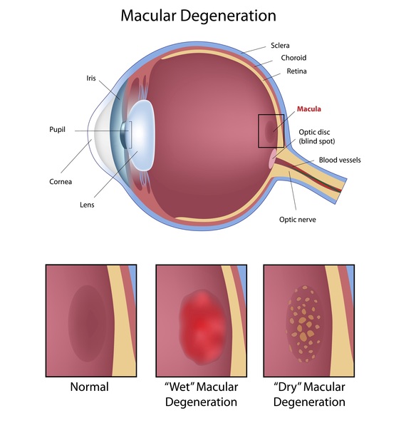
X-linked juvenile retinoschisis is a condition characterized by impaired vision that begins in childhood and occurs almost exclusively in males. This disorder affects the retina, which is a specialized light-sensitive tissue that lines the back of the eye. Damage to the retina impairs the sharpness of vision (visual acuity) in both eyes. Typically, X-linked juvenile retinoschisis affects cells in the central area of the retina called the macula. The macula is responsible for sharp central vision, which is needed for detailed tasks such as reading, driving, and recognizing faces. X-linked juvenile retinoschisis is one type of a broader disorder called macular degeneration, which disrupts the normal functioning of the macula. Occasionally, side (peripheral) vision is affected in people with X-linked juvenile retinoschisis.
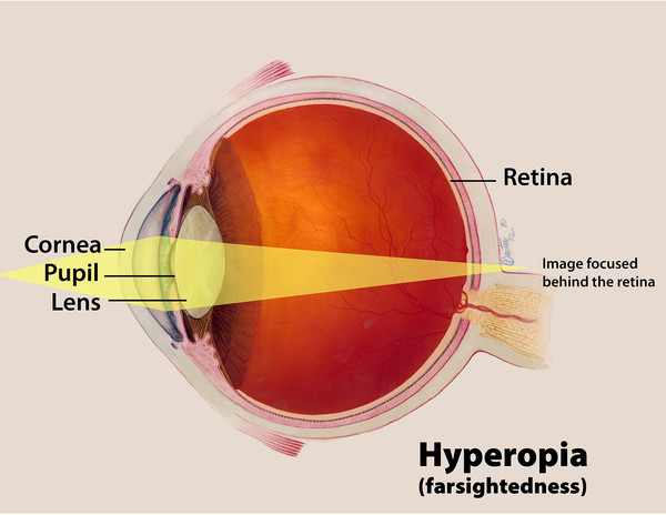
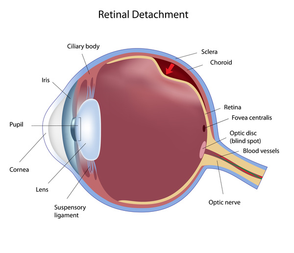
X-linked juvenile retinoschisis is usually diagnosed when affected boys start school and poor vision and difficulty with reading become apparent. In more severe cases, eye squinting and involuntary movement of the eyes (nystagmus) begin in infancy. Other early features of X-linked juvenile retinoschisis include eyes that do not look in the same direction (strabismus) and farsightedness (hyperopia). Visual acuity often declines in childhood and adolescence but then stabilizes throughout adulthood until a significant decline in visual acuity typically occurs in a man's fifties or sixties. Sometimes, severe complications develop, such as separation of the retinal layers (retinal detachment) or leakage of blood vessels in the retina (vitreous hemorrhage). These eye abnormalities can further impair vision or cause blindness.
Frequency
The prevalence of X-linked juvenile retinoschisis is estimated to be 1 in 5,000 to 25,000 men worldwide.
Causes
Mutations in the RS1 gene cause most cases of X-linked juvenile retinoschisis. The RS1 gene provides instructions for making a protein called retinoschisin, which is found in the retina. Studies suggest that retinoschisin plays a role in the development and maintenance of the retina. The protein is probably involved in the organization of cells in the retina by attaching cells together (cell adhesion).
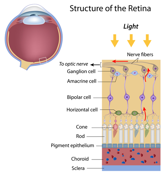
RS1 gene mutations result in a decrease in or complete loss of functional retinoschisin, which disrupts the maintenance and organization of cells in the retina. As a result, tiny splits (schisis) or tears form in the retina. This damage often forms a "spoke-wheel" pattern in the macula, which can be seen during an eye examination. In half of affected individuals, these abnormalities can occur in the area of the macula, affecting visual acuity, in the other half of cases the schisis occurs in the sides of the retina, resulting in impaired peripheral vision.
Some individuals with X-linked juvenile retinoschisis do not have a mutation in the RS1 gene. In these individuals, the cause of the disorder is unknown.
Inheritance
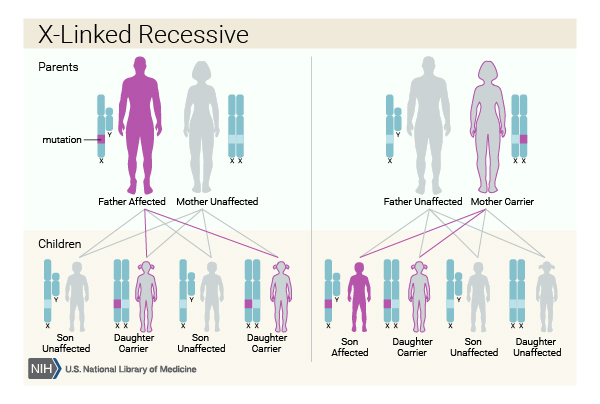
This condition is inherited in an X-linked recessive pattern. The gene associated with this condition is located on the X chromosome, which is one of the two sex chromosomes. In males (who have only one X chromosome), one altered copy of the gene in each cell is sufficient to cause the condition. In females (who have two X chromosomes), a mutation would have to occur in both copies of the gene to cause the disorder. Because it is unlikely that females will have two altered copies of this gene, males are affected by X-linked recessive disorders much more frequently than females. A characteristic of X-linked inheritance is that fathers cannot pass X-linked traits to their sons.
Other Names for This Condition
- Congenital X-linked retinoschisis
- Degenerative retinoschisis
- Juvenile retinoschisis
- X-linked retinoschisis
- XJR
Additional Information & Resources
Genetic Testing Information
Genetic and Rare Diseases Information Center
Patient Support and Advocacy Resources
Clinical Trials
Catalog of Genes and Diseases from OMIM
Scientific Articles on PubMed
References
- Apushkin MA, Fishman GA, Rajagopalan AS. Fundus findings and longitudinal study of visual acuity loss in patients with X-linked retinoschisis. Retina. 2005 Jul-Aug;25(5):612-8. doi: 10.1097/00006982-200507000-00012. Citation on PubMed
- Kim DY, Mukai S. X-linked juvenile retinoschisis (XLRS): a review of genotype-phenotype relationships. Semin Ophthalmol. 2013 Sep-Nov;28(5-6):392-6. doi: 10.3109/08820538.2013.825299. Citation on PubMed
- Molday RS, Kellner U, Weber BH. X-linked juvenile retinoschisis: clinical diagnosis, genetic analysis, and molecular mechanisms. Prog Retin Eye Res. 2012 May;31(3):195-212. doi: 10.1016/j.preteyeres.2011.12.002. Epub 2012 Jan 3. Citation on PubMed or Free article on PubMed Central
- Pimenides D, George ND, Yates JR, Bradshaw K, Roberts SA, Moore AT, Trump D. X-linked retinoschisis: clinical phenotype and RS1 genotype in 86 UK patients. J Med Genet. 2005 Jun;42(6):e35. doi: 10.1136/jmg.2004.029769. Citation on PubMed or Free article on PubMed Central
- Prenner JL, Capone A Jr, Ciaccia S, Takada Y, Sieving PA, Trese MT. Congenital X-linked retinoschisis classification system. Retina. 2006 Sep;26(7 Suppl):S61-4. doi: 10.1097/01.iae.0000244290.09499.c1. Citation on PubMed
- Yi J, Li S, Jia X, Xiao X, Wang P, Guo X, Zhang Q. Novel RS1 mutations associated with X-linked juvenile retinoschisis. Int J Mol Med. 2012 Apr;29(4):644-8. doi: 10.3892/ijmm.2012.882. Epub 2012 Jan 10. Citation on PubMed or Free article on PubMed Central
The information on this site should not be used as a substitute for professional medical care or advice. Contact a health care provider if you have questions about your health.