

Description
Pseudoachondroplasia is an inherited disorder of bone growth. It was once thought to be related to another disorder of bone growth called achondroplasia, but without that disorder's characteristic facial features. More research has demonstrated that pseudoachondroplasia is a separate disorder.
All people with pseudoachondroplasia have short stature. The average height of adult males with this condition is 120 centimeters (3 feet, 11 inches), and the average height of adult females is 116 centimeters (3 feet, 9 inches). Individuals with pseudoachondroplasia are not unusually short at birth; by the age of two, their growth rate falls below the standard growth curve.
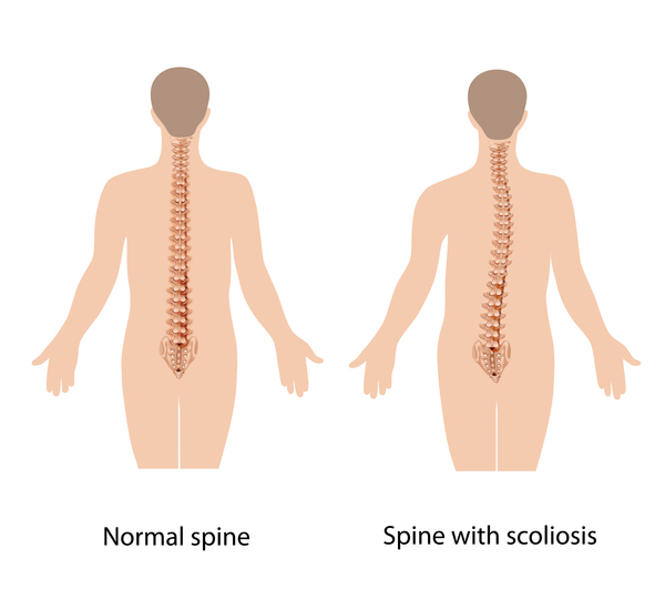
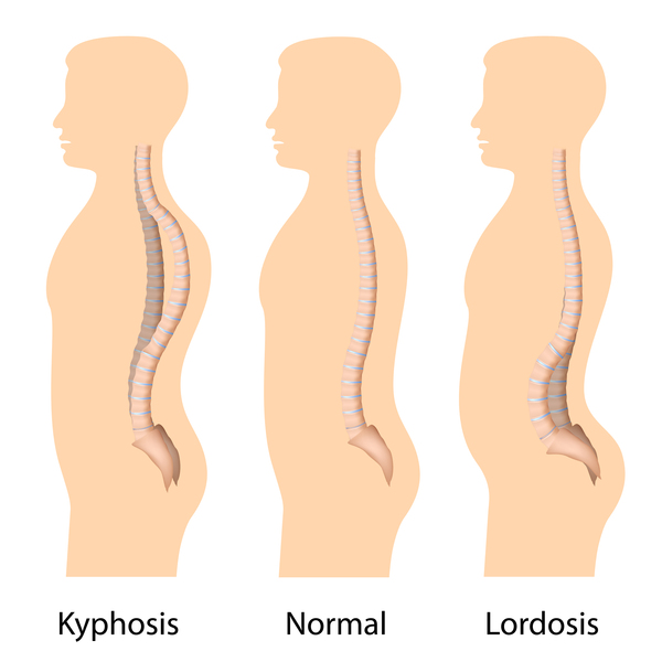
Other characteristic features of pseudoachondroplasia include short arms and legs; a waddling walk; joint pain in childhood that progresses to a joint disease known as osteoarthritis; an unusually large range of joint movement (hyperextensibility) in the hands, knees, and ankles; and a limited range of motion at the elbows and hips. Some people with pseudoachondroplasia have legs that turn outward or inward (valgus or varus deformity). Sometimes, one leg turns outward and the other inward, which is called windswept deformity. Some affected individuals have a spine that curves to the side (scoliosis) or an abnormally curved lower back (lordosis). People with pseudoachondroplasia have normal facial features, head size, and intelligence.

Frequency
The exact prevalence of pseudoachondroplasia is unknown; it is estimated to occur in 1 in 30,000 individuals.
Causes
Mutations in the COMP gene cause pseudoachondroplasia. This gene provides instructions for making a protein that is essential for the normal development of cartilage and for its conversion to bone. Cartilage is a tough, flexible tissue that makes up much of the skeleton during early development. Most cartilage is later converted to bone, except for the cartilage that continues to cover and protect the ends of bones and is present in the nose and external ears.
The COMP protein is normally found in the spaces between cartilage-forming cells called chondrocytes, where it interacts with other proteins. COMP gene mutations result in the production of an abnormal COMP protein that cannot be transported out of the cell. The abnormal protein builds up inside the chondrocyte and ultimately leads to early cell death. Early death of the chondrocytes prevents normal bone growth and causes the short stature and bone abnormalities seen in pseudoachondroplasia.
Inheritance
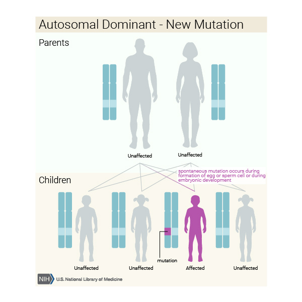
Pseudoachondroplasia is inherited in an autosomal dominant pattern, which means one copy of the altered gene in each cell is sufficient to cause the disorder.
In some cases, an affected person inherits the mutation from one affected parent. Most cases result from new mutations in the gene and occur in people with no history of the disorder in their family.

Other Names for This Condition
- PSACH
- Pseudoachondroplastic dysplasia
- Pseudoachondroplastic spondyloepiphyseal dysplasia syndrome
Additional Information & Resources
Genetic Testing Information
Genetic and Rare Diseases Information Center
Patient Support and Advocacy Resources
Clinical Trials
Catalog of Genes and Diseases from OMIM
Scientific Articles on PubMed
References
- Briggs MD, Wright MJ. COMP-Related Pseudoachondroplasia. 2004 Aug 20 [updated 2023 Nov 30]. In: Adam MP, Bick S, Mirzaa GM, Pagon RA, Wallace SE, Amemiya A, editors. GeneReviews(R) [Internet]. Seattle (WA): University of Washington, Seattle; 1993-2026. Available from http://www.ncbi.nlm.nih.gov/books/NBK1487/ Citation on PubMed
- Cao LH, Wang LB, Wang SS, Ma HW, Ji CY, Luo Y. Identification of novel and recurrent mutations in the calcium binding type III repeats of cartilage oligomeric matrix protein in patients with pseudoachondroplasia. Genet Mol Res. 2011 May 24;10(2):955-63. doi: 10.4238/vol10-2gmr1111. Citation on PubMed
- Hecht JT, Makitie O, Hayes E, Haynes R, Susic M, Montufar-Solis D, Duke PJ, Cole WG. Chondrocyte cell death and intracellular distribution of COMP and type IX collagen in the pseudoachondroplasia growth plate. J Orthop Res. 2004 Jul;22(4):759-67. doi: 10.1016/j.orthres.2003.11.010. Citation on PubMed
- Jackson GC, Mittaz-Crettol L, Taylor JA, Mortier GR, Spranger J, Zabel B, Le Merrer M, Cormier-Daire V, Hall CM, Offiah A, Wright MJ, Savarirayan R, Nishimura G, Ramsden SC, Elles R, Bonafe L, Superti-Furga A, Unger S, Zankl A, Briggs MD. Pseudoachondroplasia and multiple epiphyseal dysplasia: a 7-year comprehensive analysis of the known disease genes identify novel and recurrent mutations and provides an accurate assessment of their relative contribution. Hum Mutat. 2012 Jan;33(1):144-57. doi: 10.1002/humu.21611. Epub 2011 Oct 31. Citation on PubMed or Free article on PubMed Central
- Posey KL, Yang Y, Veerisetty AC, Sharan SK, Hecht JT. Model systems for studying skeletal dysplasias caused by TSP-5/COMP mutations. Cell Mol Life Sci. 2008 Mar;65(5):687-99. doi: 10.1007/s00018-007-7485-0. Citation on PubMed
The information on this site should not be used as a substitute for professional medical care or advice. Contact a health care provider if you have questions about your health.