

Description
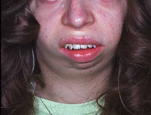
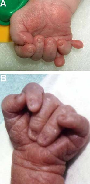
Trisomy 18, also called Edwards syndrome, is a chromosomal condition associated with abnormalities in many parts of the body. Individuals with trisomy 18 often have slow growth before birth (intrauterine growth retardation) and a low birth weight. Affected individuals may have heart defects and abnormalities of other organs that develop before birth. Other features of trisomy 18 include a small, abnormally shaped head; a small jaw and mouth; and clenched fists with overlapping fingers. Due to the presence of several life-threatening medical problems, many individuals with trisomy 18 die before birth or within their first month. Five to 10 percent of children with this condition live past their first year, and these children often have severe intellectual disability.
Frequency
Trisomy 18 occurs in about 1 in 5,000 live-born infants; it is more common in pregnancy, but many affected fetuses do not survive to term. Although women of all ages can have a child with trisomy 18, the chance of having a child with this condition increases as a woman gets older.
Causes
Most cases of trisomy 18 result from having three copies of chromosome 18 in each cell in the body instead of the usual two copies. The extra genetic material disrupts the normal course of development, causing the characteristic features of trisomy 18.
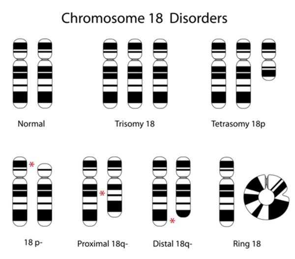
Approximately 5 percent of people with trisomy 18 have an extra copy of chromosome 18 in only some of the body's cells. In these people, the condition is called mosaic trisomy 18. The severity of mosaic trisomy 18 depends on the type and number of cells that have the extra chromosome. The development of individuals with this form of trisomy 18 may range from normal to severely affected.
Very rarely, part of the long (q) arm of chromosome 18 becomes attached (translocated) to another chromosome during the formation of reproductive cells (eggs and sperm) or very early in embryonic development. Affected individuals have two copies of chromosome 18, plus the extra material from chromosome 18 attached to another chromosome. People with this genetic change are said to have partial trisomy 18. If only part of the q arm is present in three copies, the physical signs of partial trisomy 18 may be less severe than those typically seen in trisomy 18. If the entire q arm is present in three copies, individuals may be as severely affected as if they had three full copies of chromosome 18.
Inheritance
Most cases of trisomy 18 are not inherited, but occur as random events during the formation of eggs and sperm. An error in cell division called nondisjunction results in a reproductive cell with an abnormal number of chromosomes. For example, an egg or sperm cell may gain an extra copy of chromosome 18. If one of these atypical reproductive cells contributes to the genetic makeup of a child, the child will have an extra chromosome 18 in each of the body's cells.
Mosaic trisomy 18 is also not inherited. It occurs as a random event during cell division early in embryonic development. As a result, some of the body's cells have the usual two copies of chromosome 18, and other cells have three copies of this chromosome.
Partial trisomy 18 can be inherited. An unaffected person can carry a rearrangement of genetic material between chromosome 18 and another chromosome. This rearrangement is called a balanced translocation because there is no extra material from chromosome 18. Although they do not have signs of trisomy 18, people who carry this type of balanced translocation are at an increased risk of having children with the condition.
Other Names for This Condition
- Complete trisomy 18 syndrome
- Edwards syndrome
- Trisomy 18 syndrome
- Trisomy E syndrome
Additional Information & Resources
Genetic Testing Information
Genetic and Rare Diseases Information Center
Patient Support and Advocacy Resources
Clinical Trials
Scientific Articles on PubMed
References
- Boghosian-Sell L, Mewar R, Harrison W, Shapiro RM, Zackai EH, Carey J, Davis-Keppen L, Hudgins L, Overhauser J. Molecular mapping of the Edwards syndrome phenotype to two noncontiguous regions on chromosome 18. Am J Hum Genet. 1994 Sep;55(3):476-83. Citation on PubMed or Free article on PubMed Central
- Bronsteen R, Lee W, Vettraino IM, Huang R, Comstock CH. Second-trimester sonography and trisomy 18. J Ultrasound Med. 2004 Feb;23(2):233-40. doi: 10.7863/jum.2004.23.2.233. Citation on PubMed
- Chen CP, Chern SR, Tsai FJ, Lin CY, Lin YH, Wang W. A comparison of maternal age, sex ratio and associated major anomalies among fetal trisomy 18 cases with different cell division of error. Prenat Diagn. 2005 Apr;25(4):327-30. doi: 10.1002/pd.1123. Citation on PubMed
- Crider KS, Olney RS, Cragan JD. Trisomies 13 and 18: population prevalences, characteristics, and prenatal diagnosis, metropolitan Atlanta, 1994-2003. Am J Med Genet A. 2008 Apr 1;146A(7):820-6. doi: 10.1002/ajmg.a.32200. Citation on PubMed
- Filkins K, Koos BJ. Ultrasound and fetal diagnosis. Curr Opin Obstet Gynecol. 2005 Apr;17(2):185-95. doi: 10.1097/01.gco.0000162190.83972.5a. Citation on PubMed
- Graham EM, Bradley SM, Shirali GS, Hills CB, Atz AM; Pediatric Cardiac Care Consortium. Effectiveness of cardiac surgery in trisomies 13 and 18 (from the Pediatric Cardiac Care Consortium). Am J Cardiol. 2004 Mar 15;93(6):801-3. doi: 10.1016/j.amjcard.2003.12.012. Citation on PubMed
- Moyano D, Huggon IC, Allan LD. Fetal echocardiography in trisomy 18. Arch Dis Child Fetal Neonatal Ed. 2005 Nov;90(6):F520-2. doi: 10.1136/adc.2004.070342. Epub 2005 May 24. Citation on PubMed or Free article on PubMed Central
- Pal S, Siti MI, Ankathil R, Zilfalil BA. Two cases of isochromosome 18q syndrome. Singapore Med J. 2007 May;48(5):e146-50. Citation on PubMed
- Parker SE, Mai CT, Canfield MA, Rickard R, Wang Y, Meyer RE, Anderson P, Mason CA, Collins JS, Kirby RS, Correa A; National Birth Defects Prevention Network. Updated National Birth Prevalence estimates for selected birth defects in the United States, 2004-2006. Birth Defects Res A Clin Mol Teratol. 2010 Dec;88(12):1008-16. doi: 10.1002/bdra.20735. Epub 2010 Sep 28. Citation on PubMed
- Petek E, Pertl B, Tschernigg M, Bauer M, Mayr J, Wagner K, Kroisel PM. Characterisation of a 19-year-old "long-term survivor" with Edwards syndrome. Genet Couns. 2003;14(2):239-44. Citation on PubMed
- Pont SJ, Robbins JM, Bird TM, Gibson JB, Cleves MA, Tilford JM, Aitken ME. Congenital malformations among liveborn infants with trisomies 18 and 13. Am J Med Genet A. 2006 Aug 15;140(16):1749-56. doi: 10.1002/ajmg.a.31382. Citation on PubMed
- Ramesh KH, Verma RS. Parental origin of the extra chromosome 18 in Edwards syndrome. Ann Genet. 1996;39(2):110-2. Citation on PubMed
- Rasmussen SA, Wong LY, Yang Q, May KM, Friedman JM. Population-based analyses of mortality in trisomy 13 and trisomy 18. Pediatrics. 2003 Apr;111(4 Pt 1):777-84. doi: 10.1542/peds.111.4.777. Citation on PubMed
- Tucker ME, Garringer HJ, Weaver DD. Phenotypic spectrum of mosaic trisomy 18: two new patients, a literature review, and counseling issues. Am J Med Genet A. 2007 Mar 1;143A(5):505-17. doi: 10.1002/ajmg.a.31535. Citation on PubMed
- Wapner R, Thom E, Simpson JL, Pergament E, Silver R, Filkins K, Platt L, Mahoney M, Johnson A, Hogge WA, Wilson RD, Mohide P, Hershey D, Krantz D, Zachary J, Snijders R, Greene N, Sabbagha R, MacGregor S, Hill L, Gagnon A, Hallahan T, Jackson L; First Trimester Maternal Serum Biochemistry and Fetal Nuchal Translucency Screening (BUN) Study Group. First-trimester screening for trisomies 21 and 18. N Engl J Med. 2003 Oct 9;349(15):1405-13. doi: 10.1056/NEJMoa025273. Citation on PubMed
- Yeo L, Guzman ER, Day-Salvatore D, Walters C, Chavez D, Vintzileos AM. Prenatal detection of fetal trisomy 18 through abnormal sonographic features. J Ultrasound Med. 2003 Jun;22(6):581-90; quiz 591-2. doi: 10.7863/jum.2003.22.6.581. Citation on PubMed
The information on this site should not be used as a substitute for professional medical care or advice. Contact a health care provider if you have questions about your health.