

Description
Lattice corneal dystrophy type I is an eye disorder that affects the clear, outer covering of the eye called the cornea. The cornea must remain clear for an individual to see properly; however, in lattice corneal dystrophy type I, protein clumps known as amyloid deposits cloud the cornea, which leads to vision impairment. The cornea is made up of several layers of tissue, and in lattice corneal dystrophy type I, the deposits form in the stromal layer. The amyloid deposits form as delicate, branching fibers that create a lattice pattern.
Affected individuals often have recurrent corneal erosions, which are caused by separation of particular layers of the cornea from one another. Corneal erosions are very painful and can cause sensitivity to bright light (photophobia). Lattice corneal dystrophy type I is usually bilateral, which means it affects both eyes. The condition becomes apparent in childhood or adolescence and leads to vision problems by early adulthood.
Frequency
Lattice corneal dystrophy type I is one of the most common disorders in a group of conditions that are characterized by protein deposits in the cornea (corneal dystrophies); however, it is still a rare condition. The prevalence of lattice corneal dystrophy type I is unknown.
Causes
Lattice corneal dystrophy type I is caused by mutations in the TGFBI gene. This gene provides instructions for making a protein that is found in many tissues throughout the body, including the cornea. The TGFBI protein is part of the extracellular matrix, an intricate network that forms in the spaces between cells and provides structural support to tissues. The protein is thought to play a role in the attachment of cells to one another (cell adhesion) and cell movement (migration).
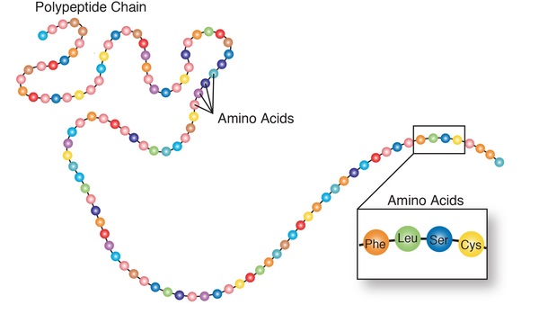
The TGFBI gene mutations involved in lattice corneal dystrophy type I change single protein building blocks (amino acids) in the TGFBI protein. Mutated TGFBI proteins abnormally clump together and form amyloid deposits. However, it is unclear how the changes caused by the gene mutations induce the protein to form deposits.
Inheritance
This condition is inherited in an autosomal dominant pattern, which means one copy of the altered gene in each cell is sufficient to cause the disorder. In most cases, an affected person has one parent with the condition.

Other Names for This Condition
- Biber-Haab-Dimmer dystrophy
Additional Information & Resources
Genetic Testing Information
Genetic and Rare Diseases Information Center
Patient Support and Advocacy Resources
Catalog of Genes and Diseases from OMIM
Scientific Articles on PubMed
References
- Kannabiran C, Klintworth GK. TGFBI gene mutations in corneal dystrophies. Hum Mutat. 2006 Jul;27(7):615-25. doi: 10.1002/humu.20334. Citation on PubMed
- Klintworth GK. Corneal dystrophies. Orphanet J Rare Dis. 2009 Feb 23;4:7. doi: 10.1186/1750-1172-4-7. Citation on PubMed or Free article on PubMed Central
- Liu Z, Wang YQ, Gong QH, Xie LX. An R124C mutation in TGFBI caused lattice corneal dystrophy type I with a variable phenotype in three Chinese families. Mol Vis. 2008 Jun 30;14:1234-9. Citation on PubMed or Free article on PubMed Central
- Munier FL, Korvatska E, Djemai A, Le Paslier D, Zografos L, Pescia G, Schorderet DF. Kerato-epithelin mutations in four 5q31-linked corneal dystrophies. Nat Genet. 1997 Mar;15(3):247-51. doi: 10.1038/ng0397-247. Citation on PubMed
- Schmitt-Bernard CF, Chavanieu A, Derancourt J, Arnaud B, Demaille JG, Calas B, Argiles A. In vitro creation of amyloid fibrils from native and Arg124Cys mutated betaIGH3((110-131)) peptides, and its relevance for lattice corneal amyloid dystrophy type I. Biochem Biophys Res Commun. 2000 Jul 5;273(2):649-53. doi: 10.1006/bbrc.2000.2955. Citation on PubMed
The information on this site should not be used as a substitute for professional medical care or advice. Contact a health care provider if you have questions about your health.