

Description
Cone-rod dystrophy is a group of related eye disorders that causes vision loss, which becomes more severe over time. These disorders affect the retina, which is the layer of light-sensitive tissue at the back of the eye. In people with cone-rod dystrophy, vision loss occurs as the light-sensing cells of the retina gradually deteriorate.
The first signs and symptoms of cone-rod dystrophy, which often occur in childhood, are usually decreased sharpness of vision (visual acuity) and increased sensitivity to light (photophobia). These features are typically followed by impaired color vision (dyschromatopsia), blind spots (scotomas) in the center of the visual field, and partial side (peripheral) vision loss. Over time, affected individuals develop night blindness and a worsening of their peripheral vision, which can limit independent mobility. Decreasing visual acuity makes reading increasingly difficult and most affected individuals are legally blind by mid-adulthood. As the condition progresses, individuals may develop involuntary eye movements (nystagmus).
There are more than 30 types of cone-rod dystrophy, which are distinguished by their genetic cause and their pattern of inheritance: autosomal recessive, autosomal dominant, and X-linked. Additionally, cone-rod dystrophy can occur alone without any other signs and symptoms or it can occur as part of a syndrome that affects multiple parts of the body.
Frequency
Cone-rod dystrophy is estimated to affect 1 in 30,000 to 40,000 individuals.
Causes
Mutations in more than 30 genes are known to cause cone-rod dystrophy. Approximately 20 of these genes are associated with the form of cone-rod dystrophy that is inherited in an autosomal recessive pattern. Mutations in the ABCA4 gene are the most common cause of autosomal recessive cone-rod dystrophy, accounting for 30 to 60 percent of cases. At least 10 genes have been associated with cone-rod dystrophy that is inherited in an autosomal dominant pattern. Mutations in the GUCY2D and CRX genes account for about half of these cases. Changes in at least two genes cause the X-linked form of the disorder, which is rare.
The genes associated with cone-rod dystrophy play essential roles in the structure and function of specialized light receptor cells (photoreceptors) in the retina. The retina contains two types of photoreceptors, rods and cones. Rods are needed for vision in low light, while cones provide vision in bright light, including color vision.
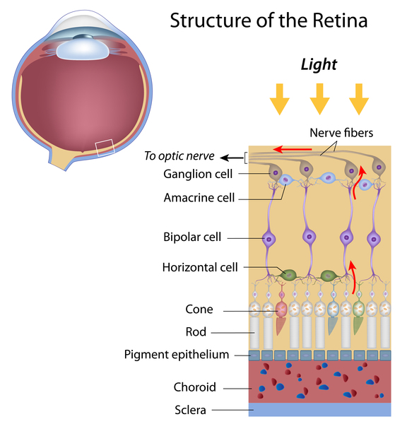

Mutations in any of the genes associated with cone-rod dystrophy lead to a gradual loss of rods and cones in the retina. The progressive degeneration of these cells causes the characteristic pattern of vision loss that occurs in people with cone-rod dystrophy. Cones typically break down before rods, which is why sensitivity to light and impaired color vision are usually the first signs of the disorder. (The order of cell breakdown is also reflected in the condition name.) Night vision is disrupted later, as rods are lost.
Some of the genes associated with cone-rod dystrophy are also associated with other eye diseases, including a group of related eye disorders called rod-cone dystrophy. Rod-cone dystrophy has signs and symptoms similar to those of cone-rod dystrophy. However, rod-cone dystrophy is characterized by deterioration of the rods first, followed by the cones, so night vision is affected before daylight and color vision. The most common form of rod-cone dystrophy is a condition called retinitis pigmentosa.
Inheritance
Cone-rod dystrophy is usually inherited in an autosomal recessive pattern, which means both copies of the gene in each cell have mutations. The parents of an individual with an autosomal recessive condition each carry one copy of the mutated gene, but they typically do not show signs and symptoms of the condition.

Less frequently, this condition is inherited in an autosomal dominant pattern, which means one copy of the altered gene in each cell is sufficient to cause the disorder. In most of these cases, an affected person has one parent with the condition.

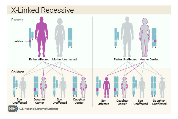
Rarely, cone-rod dystrophy is inherited in an X-linked recessive pattern. The genes associated with this form of the condition are located on the X chromosome, which is one of the two sex chromosomes. In males (who have only one X chromosome), one altered copy of the gene in each cell is sufficient to cause the condition. In females (who have two X chromosomes), a mutation would have to occur in both copies of the gene to cause the disorder. Because it is unlikely that females will have two altered copies of this gene, males are affected by X-linked recessive disorders much more frequently than females. Females with one copy of the altered gene have mild vision problems, such as decreased visual acuity. A characteristic of X-linked inheritance is that fathers cannot pass X-linked traits to their sons.
Other Names for This Condition
- Cone-rod degeneration
- Cone-rod retinal dystrophy
- CORD
- CRD
- Retinal cone-rod dystrophy
- Tapetoretinal degeneration
Additional Information & Resources
Genetic Testing Information
- Genetic Testing Registry: Cone-rod dystrophy 1

- Genetic Testing Registry: Cone-rod dystrophy 10
- Genetic Testing Registry: Cone-rod dystrophy 11
- Genetic Testing Registry: Cone-rod dystrophy 12
- Genetic Testing Registry: Cone-rod dystrophy 13
- Genetic Testing Registry: Cone-rod dystrophy 15
- Genetic Testing Registry: Cone-rod dystrophy 16
- Genetic Testing Registry: Cone-rod dystrophy 17
- Genetic Testing Registry: Cone-rod dystrophy 18
- Genetic Testing Registry: Cone-rod dystrophy 19
- Genetic Testing Registry: Cone-rod dystrophy 2
- Genetic Testing Registry: Cone-rod dystrophy 20
- Genetic Testing Registry: Cone-rod dystrophy 3
- Genetic Testing Registry: Cone-rod dystrophy 5
- Genetic Testing Registry: Cone-rod dystrophy 6
- Genetic Testing Registry: Cone-rod dystrophy 7
- Genetic Testing Registry: Cone-rod dystrophy 9
- Genetic Testing Registry: Cone-rod dystrophy
- Genetic Testing Registry: X-linked cone-rod dystrophy 1
- Genetic Testing Registry: X-linked cone-rod dystrophy 3
Genetic and Rare Diseases Information Center
Patient Support and Advocacy Resources
Clinical Trials
Catalog of Genes and Diseases from OMIM
- CONE DYSTROPHY 3; COD3
- CONE-ROD DYSTROPHY 10; CORD10
- CONE-ROD DYSTROPHY 11; CORD11
- CONE-ROD DYSTROPHY 12; CORD12
- CONE-ROD DYSTROPHY 13; CORD13
- CONE-ROD DYSTROPHY 17; CORD17
- CONE-ROD DYSTROPHY 18; CORD18
- CONE-ROD DYSTROPHY 19; CORD19
- CONE-ROD DYSTROPHY 1; CORD1
- CONE-ROD DYSTROPHY 20; CORD20
- CONE-ROD DYSTROPHY 21; CORD21
- CONE-ROD DYSTROPHY 2; CORD2
- CONE-ROD DYSTROPHY 3; CORD3
- CONE-ROD DYSTROPHY 5; CORD5
- CONE-ROD DYSTROPHY 6; CORD6
- CONE-ROD DYSTROPHY 7; CORD7
- CONE-ROD DYSTROPHY 8; CORD8
- CONE-ROD DYSTROPHY 9; CORD9
- CONE-ROD DYSTROPHY, X-LINKED, 1; CORDX1
- CONE-ROD DYSTROPHY, X-LINKED, 2; CORDX2
- CONE-ROD DYSTROPHY, X-LINKED, 3; CORDX3
Scientific Articles on PubMed
References
- Boulanger-Scemama E, El Shamieh S, Demontant V, Condroyer C, Antonio A, Michiels C, Boyard F, Saraiva JP, Letexier M, Souied E, Mohand-Said S, Sahel JA, Zeitz C, Audo I. Next-generation sequencing applied to a large French cone and cone-rod dystrophy cohort: mutation spectrum and new genotype-phenotype correlation. Orphanet J Rare Dis. 2015 Jun 24;10:85. doi: 10.1186/s13023-015-0300-3. Citation on PubMed or Free article on PubMed Central
- Hamel CP. Cone rod dystrophies. Orphanet J Rare Dis. 2007 Feb 1;2:7. doi: 10.1186/1750-1172-2-7. Citation on PubMed or Free article on PubMed Central
- Huang L, Li S, Xiao X, Jia X, Wang P, Guo X, Zhang Q. Screening for variants in 20 genes in 130 unrelated patients with cone-rod dystrophy. Mol Med Rep. 2013 Jun;7(6):1779-85. doi: 10.3892/mmr.2013.1415. Epub 2013 Apr 5. Citation on PubMed
- Huang L, Zhang Q, Li S, Guan L, Xiao X, Zhang J, Jia X, Sun W, Zhu Z, Gao Y, Yin Y, Wang P, Guo X, Wang J, Zhang Q. Exome sequencing of 47 chinese families with cone-rod dystrophy: mutations in 25 known causative genes. PLoS One. 2013 Jun 11;8(6):e65546. doi: 10.1371/journal.pone.0065546. Print 2013. Citation on PubMed or Free article on PubMed Central
- Roosing S, Thiadens AA, Hoyng CB, Klaver CC, den Hollander AI, Cremers FP. Causes and consequences of inherited cone disorders. Prog Retin Eye Res. 2014 Sep;42:1-26. doi: 10.1016/j.preteyeres.2014.05.001. Epub 2014 May 22. Citation on PubMed
- Sergouniotis PI, McKibbin M, Robson AG, Bolz HJ, De Baere E, Muller PL, Heller R, El-Asrag ME, Van Schil K, Plagnol V, Toomes C; Uk Inherited Retinal Disease Consortium; Ali M, Holder GE, Charbel Issa P, Leroy BP, Inglehearn CF, Webster AR. Disease Expression in Autosomal Recessive Retinal Dystrophy Associated With Mutations in the DRAM2 Gene. Invest Ophthalmol Vis Sci. 2015 Dec;56(13):8083-90. doi: 10.1167/iovs.15-17604. Citation on PubMed
- Thiadens AA, Phan TM, Zekveld-Vroon RC, Leroy BP, van den Born LI, Hoyng CB, Klaver CC; Writing Committee for the Cone Disorders Study Group Consortium; Roosing S, Pott JW, van Schooneveld MJ, van Moll-Ramirez N, van Genderen MM, Boon CJ, den Hollander AI, Bergen AA, De Baere E, Cremers FP, Lotery AJ. Clinical course, genetic etiology, and visual outcome in cone and cone-rod dystrophy. Ophthalmology. 2012 Apr;119(4):819-26. doi: 10.1016/j.ophtha.2011.10.011. Epub 2012 Jan 20. Citation on PubMed
The information on this site should not be used as a substitute for professional medical care or advice. Contact a health care provider if you have questions about your health.