



Description
Color vision deficiency (sometimes called color blindness) represents a group of conditions that affect the ability to see certain colors. Color vision deficiency can be present from birth (congenital) or it can appear later in life as the result of an injury or other visual impairment (acquired).
There are four main forms of color vision deficiency:
- Red-green color vision deficiency
- Blue-yellow color vision deficiency
- Blue cone monochromacy
- Complete color vision deficiency
Red-green color vision deficiency is the most common form of color vision deficiency. Affected individuals have trouble distinguishing between some shades of red and green.
Red-green color vision deficiency can be further divided into four subtypes:
- Protanomaly: reds appear less vibrant
- Deuteranomaly: greens appear as blues or yellows, and most other colors are less vibrant
- Protanopia: red cannot be perceived, dark colors look orange or green, and most colors look blue-ish or gold-ish
- Deuteranopia: green cannot be perceived, it is difficult to distinguish between red and green, and most colors look blue-ish or gold-ish
People with blue-yellow color vision deficiency have problems differentiating shades of blue and green and difficulty distinguishing between dark blue and black.
Blue-yellow color vision deficiency can be further divided into two subtypes:
- Tritanopia: blue cannot be perceived, and most colors look red-ish or purple-ish
- Tritanomaly: blues appear as green, yellows are difficult to perceive
Color vision deficiency that affects red-green or blue-yellow color vision disrupts color perception but usually does not affect the sharpness of vision (visual acuity). An exception to this is Bornholm eye disease, which often causes red-green color vision deficiency, low visual acuity, nearsightedness (myopia), and additional vision problems.
Blue cone monochromacy and complete color vision deficiency allow for little or no color vision. Affected individuals often have vision impairment and additional vision problems, which can include increased sensitivity to light (photophobia), involuntary back-and-forth eye movements (nystagmus), and myopia. Complete color vision deficiency is called rod monochromacy or achromatopsia.
Frequency
Color vision deficiency is estimated to affect about 2.5 percent of individuals worldwide. This condition is more common in men than in women.
Red-green color vision deficiency is the most common form of color vision deficiency. Among populations with Northern European ancestry, red-green color vision deficiency occurs in about 1 in 12 men and 1 in 200 women. Red-green color vision deficiency is also common in East Asian populations. This condition is less common in almost all other populations that have been studied.
Blue-yellow color vision deficiency affects men and women equally. This type occurs in fewer than 1 in 10,000 people worldwide.
Blue cone monochromacy and complete color vision deficiency are rare. Complete color vision deficiency affects men and women equally.
Causes
Genetic changes that cause disease are called pathogenic variants. Pathogenic variants in several different genes can cause color vision deficiency. Variants in the OPN1LW, OPN1MW, and OPN1SW genes are common causes of color vision deficiency.
Pathogenic variants in the OPN1LW and OPN1MW genes cause red-green color vision deficiency, Bornholm eye disease, and blue cone monochromacy. Pathogenic variants in the OPN1SW genes cause blue-yellow color vision deficiency. Variants in several other genes cause achromatopsia.
The OPN1LW, OPN1MW, and OPN1SW genes provide instructions for making proteins called opsins that play essential roles in color vision. Opsins act as color pigments in the retina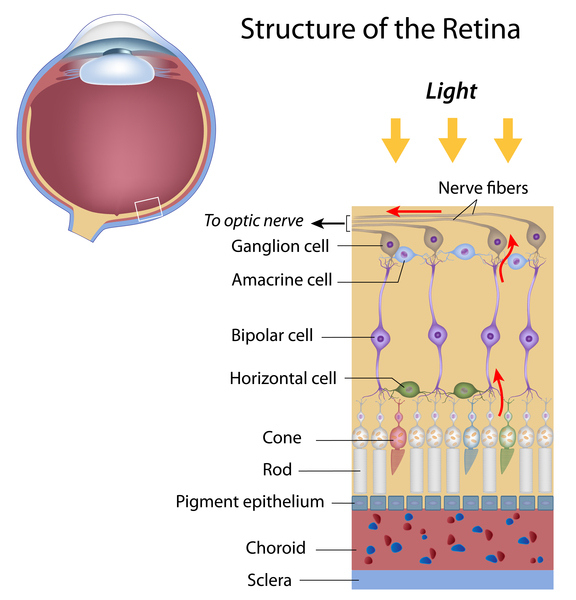 , which is the light-sensitive tissue at the back of the eye
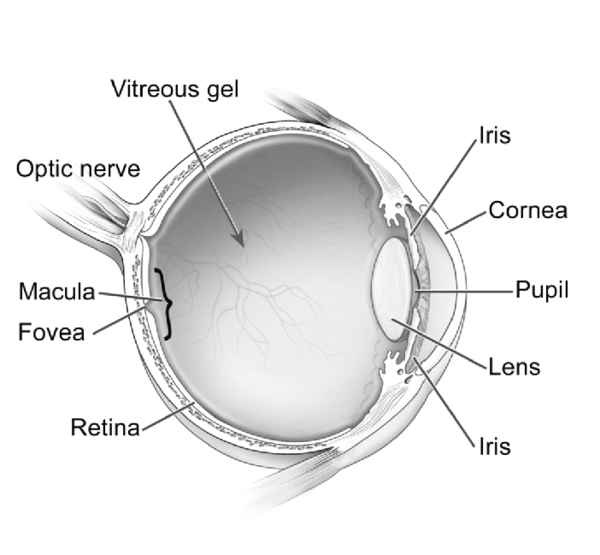 . The retina contains two types of light receptor cells, called rods and cones
 , that transmit visual signals from the eye to the brain. Rods provide vision in low light. Cones provide vision in bright light, including color vision and high visual acuity. There are three types of cones, each containing a specific opsin that is most sensitive to a particular wavelength of light. The retina and the brain combine input from all three types of cones to produce normal color vision.
The opsin made from the OPN1LW gene is more sensitive to light in the red part of the visible spectrum (long-wavelength light), and cones with this opsin are called long-wavelength-sensitive cones or L cones. The opsin made from the OPN1MW gene is more sensitive to light in the middle of the visible spectrum (green light), and cones with this opsin are called middle-wavelength-sensitive cones or M cones. The opsin made from the OPN1SW gene is more sensitive to light in the blue part of the visible spectrum (short-wavelength light), and cones with this opsin are called short-wavelength-sensitive cones or S cones.
Pathogenic variants in the OPN1LW or OPN1MW gene can cause cells to produce abnormal versions of the opsins for the L or M cones, or they can prevent these cells from producing opsins at all. When either the L or M cone is affected, red-green color vision is impaired. When changes in these genes prevent both L and M cones from functioning normally, and only S cones are functional, the result is blue cone monochromancy.
Pathogenic variants in the OPN1SW gene can prevent S cones from producing opsins that function properly. This can alter the perception of the color blue. As a result, it is difficult or impossible to detect differences between shades of blue and green, and affected individuals have problems distinguishing between dark blue and black.
Acquired color vision deficiency is not caused by gene variants. It can be caused by other eye disorders, certain medications, or environmental exposures. It can also be a symptom of other conditions. Color vision may also decline with age.
Inheritance
Color vision deficiency can have different inheritance patterns depending on the gene involved.
Red-green color vision deficiency and blue cone monochromacy are inherited in an X-linked pattern. The OPN1LW and OPN1MW genes are located on the X chromosome, which is one of the two sex chromosomes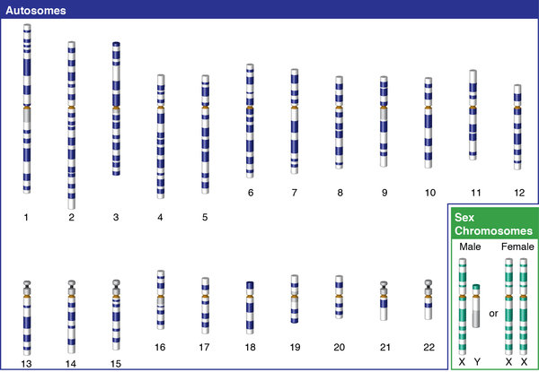 . In males (who have only one X chromosome), a pathogenic variant in the only copy of the gene in each cell is sufficient to cause the condition. In females (who have two X chromosomes), two altered copies of the gene are needed to cause the disorder. Because it is unlikely that females will have two altered copies, males are affected by red-green color vision deficiency and blue cone monochromacy much more frequently than females. A characteristic of X-linked inheritance is that fathers cannot pass X-linked traits to their sons.
Blue-yellow color vision deficiency is inherited in an autosomal dominant 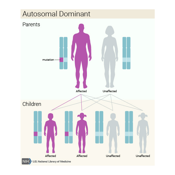 pattern, which means one copy of the altered OPN1SW gene in each cell is sufficient to cause the condition. In many cases, an affected person inherits the condition from an affected parent.
Other Names for This Condition
- Color blindness
- Color vision defects
- Defective color vision
Additional Information & Resources
Genetic Testing Information

Genetic and Rare Diseases Information Center
Patient Support and Advocacy Resources
Clinical Trials
Catalog of Genes and Diseases from OMIM
Scientific Articles on PubMed
References
- Almustanyir A. A Global Perspective of Color Vision Deficiency: Awareness, Diagnosis, and Lived Experiences. Healthcare (Basel). 2025 Aug 17;13(16):2031. doi: 10.3390/healthcare13162031. Citation on PubMed
- Deeb SS. Molecular genetics of color-vision deficiencies. Vis Neurosci. 2004 May-Jun;21(3):191-6. doi: 10.1017/s0952523804213244. Citation on PubMed
- Deeb SS. The molecular basis of variation in human color vision. Clin Genet. 2005 May;67(5):369-77. doi: 10.1111/j.1399-0004.2004.00343.x. Citation on PubMed
- Fayaz F, Jafarzadehpur E, Ahmad MA, Amini Vishteh R, Pishnamaz MR, Hoseinzade MS, Mirzaee Saba L, Kamali M, Yekta A, Khabazkhoob M. Factors affecting color vision: a systematic review. Int J Ophthalmol. 2026 Jul 18;19(7):1395-1405. doi: 10.18240/ijo.2026.07.21. eCollection 2026. Citation on PubMed
- Gardner JC, Michaelides M, Holder GE, Kanuga N, Webb TR, Mollon JD, Moore AT, Hardcastle AJ. Blue cone monochromacy: causative mutations and associated phenotypes. Mol Vis. 2009;15:876-84. Epub 2009 May 1. Citation on PubMed or Free article on PubMed Central
- Jeong YD, Cho J, Son Y, Jo Y, Yim Y, Kim TH, Kim S, Cho H, Rahmati M, Smith L, Woo HG, Kim JH, Kim YJ, Yang JM, Yon DK. Global Prevalence of Congenital Color Vision Deficiency among Children and Adolescents, 1932-2022. Ophthalmology. 2025 Dec;132(12):1431-1444. doi: 10.1016/j.ophtha.2025.07.031. Epub 2025 Aug 5. Citation on PubMed
- Kohl S, Jagle H, Wissinger B, Zobor D. Achromatopsia. 2004 Jun 24 [updated 2018 Sep 20]. In: Adam MP, Bick S, Mirzaa GM, Wallace SE, Amemiya A, editors. GeneReviews(R) [Internet]. Seattle (WA): University of Washington, Seattle; 1993-2026. Available from http://www.ncbi.nlm.nih.gov/books/NBK1418/ Citation on PubMed
- Neitz J, Neitz M. The genetics of normal and defective color vision. Vision Res. 2011 Apr 13;51(7):633-51. doi: 10.1016/j.visres.2010.12.002. Epub 2010 Dec 15. Citation on PubMed or Free article on PubMed Central
- Pasmanter N, Munakomi S. Physiology, Color Perception. 2022 Sep 12. In: StatPearls [Internet]. Treasure Island (FL): StatPearls Publishing; 2022 Jan-. Available from http://www.ncbi.nlm.nih.gov/books/NBK544355/ Citation on PubMed
The information on this site should not be used as a substitute for professional medical care or advice. Contact a health care provider if you have questions about your health.