

Description
Osteogenesis imperfecta (OI) is a group of genetic disorders that mainly affect the bones. The term "osteogenesis imperfecta" means imperfect bone formation. People with this condition have bones that break (fracture) easily, often from mild trauma or with no apparent cause. Multiple fractures are common, and in severe cases, can occur even before birth. Milder cases may involve only a few fractures over a person's lifetime.
There are at least 19 recognized forms of osteogenesis imperfecta, designated type I through type XIX. Several types are distinguished by their signs and symptoms, although their characteristic features overlap. Increasingly, genetic causes are used to define rarer forms of osteogenesis imperfecta. Type I (also known as classic non-deforming osteogenesis imperfecta with blue sclerae) is the mildest form of osteogenesis imperfecta. Type II (also known as perinatally lethal osteogenesis imperfecta) is the most severe. Other types of this condition, including types III (progressively deforming osteogenesis imperfecta) and IV (common variable osteogenesis imperfecta with normal sclerae), have signs and symptoms that fall somewhere between these two extremes.
The milder forms of osteogenesis imperfecta, including type I, are characterized by bone fractures during childhood and adolescence that often result from minor trauma, such as falling while learning to walk. Fractures occur less frequently in adulthood. People with mild forms of the condition typically have a blue or grey tint to the part of the eye that is usually white (the sclera), and about half develop hearing loss in adulthood. Unlike more severely affected individuals, people with type I are usually of normal or near normal height.
Other types of osteogenesis imperfecta are more severe, causing frequent bone fractures that are present at birth and result from little or no trauma. Additional features of these types can include blue sclerae of the eyes, short stature, curvature of the spine (scoliosis), joint deformities (contractures), hearing loss, respiratory problems, and a disorder of tooth development called dentinogenesis imperfecta. Mobility can be reduced in affected individuals, and some may use a walker or wheelchair. The most severe forms of osteogenesis imperfecta, particularly type II, can include an abnormally small, fragile rib cage and underdeveloped lungs. Infants with these abnormalities may have life-threatening problems with breathing and can die shortly after birth.
Frequency
Osteogenesis imperfecta affects approximately 1 in 10,000 to 20,000 people worldwide. An estimated 25,000 to 50,000 people in the United States have the condition.
Causes
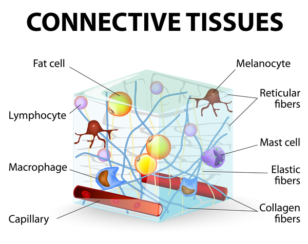
Osteogenesis imperfecta can be caused by mutations in one of several genes. Mutations in the COL1A1 and COL1A2 genes cause approximately 90 percent of all cases. These genes provide instructions for making proteins that are used to assemble type I collagen. This type of collagen is the most abundant protein in bone, skin, and other connective tissues that provide structure and strength to the body.
Osteogenesis imperfecta type I is caused by mutations in the COL1A1 gene or, less commonly, the COL1A2 gene. These genetic changes reduce the amount of type I collagen produced in the body, though the molecules that are produced are normal. A reduction in type I collagen causes bones to be brittle and to fracture easily. The mutations that cause osteogenesis imperfecta types II, III, and IV occur in either the COL1A1 or COL1A2 gene. These mutations typically alter the structure of type I collagen molecules, resulting in abnormal type I collagen. A defect in the structure of type I collagen weakens connective tissues, particularly bone, resulting in the characteristic features of these more severe types of osteogenesis imperfecta.
Mutations in other genes cause rare forms of osteogenesis imperfecta. Many of these genes provide instructions for proteins that help process type I collagen into its mature form. Mutations in these genes disrupt different steps in the production of collagen molecules. These changes weaken connective tissues, leading to severe bone abnormalities and problems with growth. Other genes involved in osteogenesis imperfecta provide instructions for making proteins that control the development and function of bone-forming cells. Mutations in these genes impair normal bone development, causing the bones to be brittle and to fracture easily.
Inheritance
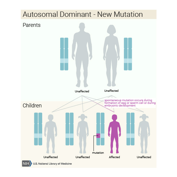
When caused by mutations in the COL1A1 or COL1A2 gene, osteogenesis imperfecta has an autosomal dominant pattern of inheritance, which means one copy of the altered gene in each cell is sufficient to cause the condition. Many people with type I or type IV osteogenesis imperfecta inherit a mutation from a parent who has the disorder. Most infants with more severe forms of osteogenesis imperfecta (such as type II and type III) have no history of the condition in their family. In these infants, the condition is caused by new (sporadic) mutations in the COL1A1 or COL1A2 gene. Type V is also inherited in an autosomal dominant pattern.

Less commonly, osteogenesis imperfecta has an autosomal recessive pattern of inheritance. Autosomal recessive inheritance means two copies of the gene in each cell are altered. The parents of a child with an autosomal recessive disorder typically are not affected, but each carry one copy of the altered gene. Types VI through XVIII follow this pattern of inheritance.
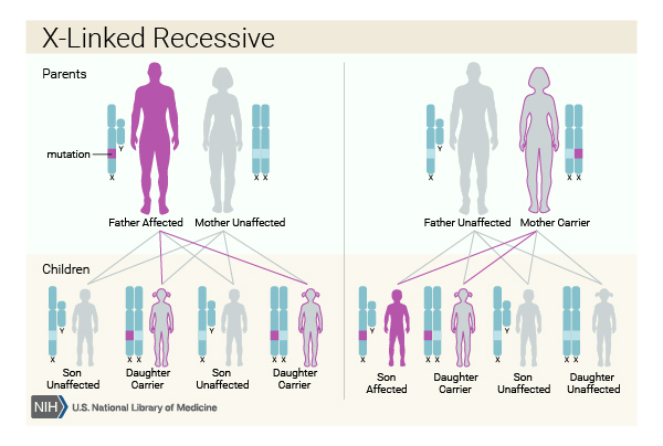
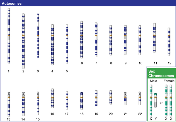
Osteogenesis imperfecta type XIX is inherited in an X-linked recessive pattern. A condition is considered X-linked if the mutated gene that causes the disorder is located on the X chromosome, one of the two sex chromosomes in each cell. In males, who have only one X chromosome, a mutation in the only copy of the gene in each cell is sufficient to cause the condition. In females (who have two X chromosomes), a mutation would have to occur in both copies of the gene to cause the disorder. Because it is unlikely that females will have two altered copies of this gene, males are affected by X-linked recessive disorders much more frequently than females. A characteristic of X-linked inheritance is that fathers cannot pass X-linked traits to their sons.
Other Names for This Condition
- Brittle bone disease
- Fragilitas ossium
- OI
- Vrolik disease
Additional Information & Resources
Genetic Testing Information
- Genetic Testing Registry: Osteogenesis imperfecta type 11

- Genetic Testing Registry: Osteogenesis imperfecta type 13
- Genetic Testing Registry: Osteogenesis imperfecta type 14
- Genetic Testing Registry: Osteogenesis imperfecta type 15
- Genetic Testing Registry: Osteogenesis imperfecta type 16
- Genetic Testing Registry: Osteogenesis imperfecta type 17
- Genetic Testing Registry: Osteogenesis imperfecta type 6
- Genetic Testing Registry: Osteogenesis imperfecta
- Genetic Testing Registry: Osteogenesis imperfecta type 10
- Genetic Testing Registry: Osteogenesis imperfecta type 12
- Genetic Testing Registry: Osteogenesis imperfecta type 5
- Genetic Testing Registry: Osteogenesis imperfecta type 7
- Genetic Testing Registry: Osteogenesis imperfecta type 8
- Genetic Testing Registry: Osteogenesis imperfecta type 9
- Genetic Testing Registry: Osteogenesis imperfecta type I
- Genetic Testing Registry: Osteogenesis imperfecta type III
- Genetic Testing Registry: Osteogenesis imperfecta with normal sclerae, dominant form
- Genetic Testing Registry: Osteogenesis imperfecta, type 18
- Genetic Testing Registry: Osteogenesis imperfecta, type 19
Genetic and Rare Diseases Information Center
Patient Support and Advocacy Resources
Clinical Trials
Catalog of Genes and Diseases from OMIM
- OSTEOGENESIS IMPERFECTA, TYPE I; OI1
- OSTEOGENESIS IMPERFECTA, TYPE IV; OI4
- OSTEOGENESIS IMPERFECTA, TYPE XIX; OI19
- OSTEOGENESIS IMPERFECTA, TYPE III; OI3
- OSTEOGENESIS IMPERFECTA, TYPE IX; OI9
- OSTEOGENESIS IMPERFECTA, TYPE II; OI2
- OSTEOGENESIS IMPERFECTA, TYPE VII; OI7
- OSTEOGENESIS IMPERFECTA, TYPE V; OI5
- OSTEOGENESIS IMPERFECTA, TYPE XI; OI11
- OSTEOGENESIS IMPERFECTA, TYPE VIII; OI8
- OSTEOGENESIS IMPERFECTA, TYPE XVIII; OI18
- OSTEOGENESIS IMPERFECTA, TYPE XIII; OI13
- OSTEOGENESIS IMPERFECTA, TYPE XIV; OI14
- OSTEOGENESIS IMPERFECTA, TYPE XV; OI15
- OSTEOGENESIS IMPERFECTA, TYPE X; OI10
- OSTEOGENESIS IMPERFECTA, TYPE XII; OI12
- OSTEOGENESIS IMPERFECTA, TYPE XVII; OI17
- OSTEOGENESIS IMPERFECTA, TYPE VI; OI6
- OSTEOGENESIS IMPERFECTA, TYPE XVI; OI16
Scientific Articles on PubMed
References
- Byers PH, Pyott SM. Recessively inherited forms of osteogenesis imperfecta. Annu Rev Genet. 2012;46:475-97. doi: 10.1146/annurev-genet-110711-155608. Citation on PubMed
- Kang H, Aryal A C S, Marini JC. Osteogenesis imperfecta: new genes reveal novel mechanisms in bone dysplasia. Transl Res. 2017 Mar;181:27-48. doi: 10.1016/j.trsl.2016.11.005. Epub 2016 Nov 19. Citation on PubMed
- Lim J, Grafe I, Alexander S, Lee B. Genetic causes and mechanisms of Osteogenesis Imperfecta. Bone. 2017 Sep;102:40-49. doi: 10.1016/j.bone.2017.02.004. Epub 2017 Feb 15. Citation on PubMed or Free article on PubMed Central
- Marini JC, Forlino A, Bachinger HP, Bishop NJ, Byers PH, Paepe A, Fassier F, Fratzl-Zelman N, Kozloff KM, Krakow D, Montpetit K, Semler O. Osteogenesis imperfecta. Nat Rev Dis Primers. 2017 Aug 18;3:17052. doi: 10.1038/nrdp.2017.52. Citation on PubMed
- Steiner RD, Basel D. COL1A1/2 Osteogenesis Imperfecta. 2005 Jan 28 [updated 2024 Mar 14]. In: Adam MP, Feldman J, Mirzaa GM, Pagon RA, Wallace SE, Bean LJH, Gripp KW, Amemiya A, editors. GeneReviews(R) [Internet]. Seattle (WA): University of Washington, Seattle; 1993-2024. Available from http://www.ncbi.nlm.nih.gov/books/NBK1295/ Citation on PubMed
- Tournis S, Dede AD. Osteogenesis imperfecta - A clinical update. Metabolism. 2018 Mar;80:27-37. doi: 10.1016/j.metabol.2017.06.001. Epub 2017 Jun 8. Citation on PubMed
The information on this site should not be used as a substitute for professional medical care or advice. Contact a health care provider if you have questions about your health.